Vegetative Growth Simplistically, wood fungi live through two functionally different phases: the vegetative stage for mycelial spread and the reproductive stage for the elab-oration of spore-producing structures. Rayner et al. (1985) extended the development of a fungus in arrival, establishment, exploitation, and exit. The vegetative, asexual stage consists in wood fungi of vegetative hyphae with some specialized forms. The reproductive stage can both occur asexually or sexually (Schwantes 1996).
Functional specialization of the mycelium occurs during the vegetative stage: germination, infection, spread, and survival. These functions are correlated with different "fungal organs". Spores (conidia, chlamydospores, also the sex-ually derived asco- and basidiospores) germinate under suitable conditions (moisture, temperature).
The young germ hypha first shows some nuclei be-fore the young mycelium grows with septation in the monokaryotic condi( ion. N1ycelial growth takes place via mitoses and synthesis of hyphal biomass. in-fection and colonization of new substrates occurs by spores, hyphae, mycelium, and special forms like bore-hyphae, transpressoria, strands, and rhizornorphs. Prerequisites for the colonization of a substrate are suitable humidity and nu-trient availability in the substrate or, like in Serpula lacrymans, the ability of a fungus to transport nutrients and water and last, whether and by which organisms the substrate is already occupied (Rayner and Boddy 1988).
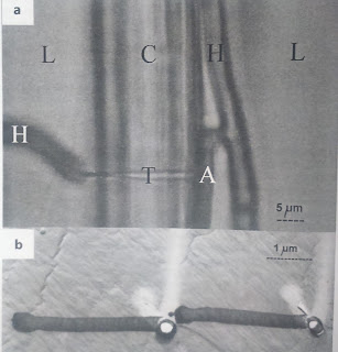
Boring microhyphae of 0.1-0.4 iirn diameter develop e.g., in Phellinus pini at the hvphal tip without recognizable septum and produce boreholes of 0.3 —3.3 p.m diameter probably by enzyme action (Schmid and Liese 1966). The appresso-rium is a hypha for the mechanical fixation to the substrate (Fig. 2.5a). The transpressorium (Fig. 2.5b) of the blue-stain fungi (Chap. 6.2) is a special-ized boring hypha (Liese 1970); it is still unknown whether the penetration of the woody cell wall is by mechanical and/or enzymatic action. Transpres-soria have also been found in the white-rot fungus Phellinus pini (Liese and Schmid 1966).
Strands (cords) (Fig.) develop in a number of house-rot fungi and usually consist of three hyphal types, vegetative hyphae, thin fiber (skeletal) hyphae with mostly thick walls for strengthening, and broad vessel hyphae for nutrient transport (Nuss et al. 1991). These hyphae form a distinct mycelium in the lon-gitudinal direction, which is, however, not so well organized like the tissue-like structure of the rhizomorphs. Also in contrast to rhizomorphs, strands develop behind the mycelial growth front. Particularly Serpula lacrymans overgrows larger distances of non-woody substrates and penetrates through masonry
Serpula lacrymans
Strands white, silver-grey to brown, more than 5 mm to 3 cm diameter, separable, wit flabby mycelium in between, thick strands when dry breaking with clearly audible cracking (strands with mold contamination often not cracking any more), often in masonry; (S. himantioides: strands thinner than 2 mm and fibers 2-3.5 pm in diameter)
Vegetative hyphae hyaline, partly yellowish, with large clamp connections, 2-4 pm in diameter Vessels at least partly numerous (in groups), 5-60 pm in diameter, not or rarely branched, with bar thickening up to 13 p.m high Fibers refractive, 3-5 pm diameter, straight-lined, stiff, septa not visible, no clamps, lumens often visible Coniophora puteana, C. marmorata Strands first bright, then brown to black, up to 2 mm wide, to 1 mm thick, root-like, hardly removable (not so in C. marmorata), when removed usually fragile, partly with brighter center, underlying wood becoming black, also in masonry Vegetative hyphae usually without clamps, rarely multiple clamps (often indistinct when branched), 2-6 pm in diameter Vessels surrounded and interwoven by many fine hyphae (0.5-1.5 pm in diameter), difficult to isolate (preparation with KOH solution); drop-shaped, hyaline to brownish secretions (1-5 pm in diameter) often on hyphae; vessels due to preparation irregularly formed or distorted, up to 30 pm in diameter, thin-walled (slightly thick-walled with C. marmorata), without bars, with septa Fibers pale to dark brown, 2-4 pm in diameter, somewhat thick-walled, with relatively broad, usually visible lumen, some also branched, to be confused with generative hyphae Antrodia vaillantii, A. serialis, A. sinuosa, A. xantha Strands white to cream, partly somewhat yellowing or infected by molds, also ice flower-like, flexible also when dry, up to 7 mm in diameter, possibly also within masonry Vegetative hyphae with few clamps, 2-4 pm in diameter, often somewhat thick-walled; in KOH somewhat swelling Vessels not rare but in old strands difficult to isolate, up to 25 pm in diameter, thick-wal with middle lumen, without bars Fibers hyaline (in Antrodia xantha partly somewhat yellowish), numerous, 2-4 pm in diameter, hyphal tips with tapering ending cell walls, straight-lined, mostly unbranched, insoluble in KOH, but partly somewhat swelling, then with "blown up" hyphal segments
20 strand-forming wood decay fungi based on Huckfeldt and Schmidt (200- 2006) is given in Appendix 1. The rhizomorphs ofArmillaria mellea (Fig. 2.7) are tissue-like mycelial bun dies with apical growth and consist of a black, gelatinous bark layer, followei by a pseudoparenchyma, and a central, loosely interwoven pith with vessel any

Comments